ABR (auditory brainstem response) is mainly used for:
To
It can be combined with other audiology tests to identify the nature of hearing loss; it is most commonly used to check for post-cochlear lesions: such as prolonged latency of each wave, prolonged wave interval, significant difference in latency or wave interval between ears, and waveform differentiation The difference indicates the possibility of post-cochlear lesions.
The main differential diagnosis of ABR:
①Transmissive deafness: The V-wave response threshold is increased but the threshold latency is in the normal range. The acoustic latency-intensity function curve shifts to the right.
② Meniere's disease: Deafness with reinstatement is manifested as an increase in the V-wave threshold, but when the acoustic stimulation is within 20dB above the threshold, the incubation period is shortened and reaches a normal value;
③Acoustic neuroma: I-V wave interval is prolonged or V wave disappears, but if the patient's I wave is not clear, the false positive rate is very high. At this time, the comprehensive analysis of cochlear electrogram should be combined to improve the diagnostic accuracy. The difference between the I-V interval between the two ears is greater than 0.4ms, or the I-V interval on one side is greater than 4.6ms (age and gender factors should be considered), it indicates that there is a posterior cochlear disease;
④Diagnosis of brainstem lesions: Multiple sclerosis, brainstem vascular lesions and brainstem tumors can also cause the amplitude of the evoked potential to decrease, the incubation period to prolong, or the waveform to disappear. It should be identified based on the medical history and related examinations. Functional deafness and pseudo-deafness: The hearing threshold can be assessed objectively, but it should be noted that short-latency potentials and short-sound examinations can easily underestimate residual hearing in the low-frequency domain.
BAEP (auditory evoked potential) is mainly used for:
Audiometry in otology (pediatric hearing screening, functional and organic deafness), auditory pathway nerves and brainstem functional or organic diseases (peripheral acoustic neuropathy, acoustic neuroma, brainstem intramedullary tumor, multiple sclerosis, Brainstem vascular disease, genetic metabolic and degenerative diseases, medullary cavitation), chronic viral infection and meningitis, encephalitis, epilepsy, tinnitus, sequelae of brain oscillation, hysteria and deafness, coma and brain death, hepatic encephalopathy, chronic Renal failure, diabetes, alcohol and smokers, and anesthesia.
Basic parameters
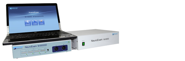
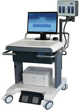
A. System composition: preamplifier, stimulation system, data processing system, trolley, power supply system and accessories
B. Trolley size (length X width X height): 750mm x 700mm x 1150mm
C. Continuous working time: ≥4 hours
To
2. Amplifier
A. Number of channels: 4 channels with retractable arm
*b. Input impedance: ≥2000 megohms
*c. Input short-circuit noise: ≤0.4μVrms (0.1Hz-10kHz)
*d. Common mode rejection ratio: ≥115dB
e. Sensitivity: 0.01μV/div-20mv/div 1mS/div-500mS/div
F. Filtering frequency: 0.1Hz-20KHz;
g. Transmission mode: photoelectric isolation, AC power supply
h. Input signal range: 1μV – 10mV
To
3. Recorder
a. Interface technology: USB
b. A/D conversion rate: 16Bit
c. Sampling rate: 200kHz (50kHz/channel)
d. Scanning time: 1ms-6s
e. Stacking times: 1-4000 times (safety limit: up to 4000 times)
f. Maximum time for collecting data: unlimited time
To
4. Stimulator
a. Acoustic stimulator:
A1, dual-channel output interface, can choose unilateral or bilateral simultaneous output
A2. Stimulation frequency: 0.05 Hz-50Hz
A3. Stimulus intensity: 0-120dB SPL
A4. Pure audio frequency: 0.5-8khz
A5. Polarity: sparse wave, dense wave, alternating sparse wave;
A6. Stimulus type: short tone, short tone


