Welcome to the website of Henan Anhel Medical Equipment Co., Ltd.!
- Molecular diagnosis
- Rapid Tests
- Drug of Abuse
-
Immunofluorescence
- Urinalysis Tests
-
POCT System
-
ELISA Test
-
DOT Test
-
Chemistry
- Ultrasonic
- Hemodialysis
- Ventilator
- ECG/EEG/EMG
-
Dermatology
- AN-specific electromagnetic wave therapy device
- AN-KL Carbon Dioxide Laser Treatment Machine (Scanning)
- AN high-energy narrow-band red and blue light therapy system
- AN Steam Therapy Fumigation Apparatus
- AN-308nm Excimer Light Skin Treatment System
- AN-5000 308 Home Excimer Phototherapy Apparatus
- AN-LED Home Spectral Therapy Apparatus
- AN 311 Ultraviolet Light Therapy Apparatus
- AN LED Spectrum Therapy Apparatus
- AN Medical Video Wood Lamp
- CT/MR
-
Ophthalmology Department
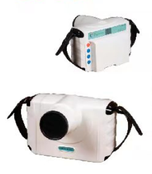
- AN-BLX5 Dental X-Ray
- AN-BLX10 Dental X-Ray
- AN-BLX8P High Frequency Dental X-Ray
- AN-BLX9 Dental X-Ray
- AN-BLX6 Dental X-Ray
- AN-BLX8 Dental X-Ray
- AN-digital oral X-ray imaging system HDR-500/600
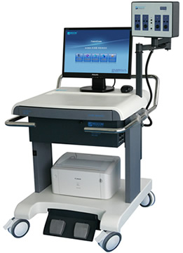
- AN-Visual evoked potential meter M-800E
- AN-AOV-FB Excimer Laser Ophthalmology Treatment Machine
- AN-YZ3 Handheld Slit Lamp Microscope
- Dental
- Hematology Analyzer
- Chemistry Analyzer
-
POCT SYSTEM
- AN-AT1 Touch Screen HD Black and White Ultra
- AN-RS-N50 full digital notebook ultrasound diagnostic instru
- AN-RS-N50 (VET) all-digital notebook ultrasound diagnostic i
- AN-A6 All-digital veterinary B-ultrasound diagnostic instrum
- AN-M6 Veterinary B Ultrasound Diagnostic Instrument
- AN-A10 All-digital veterinary B-ultrasound diagnostic instru
- AN-M10 Veterinary B Ultrasound Diagnostic Instrument


