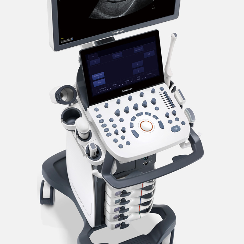
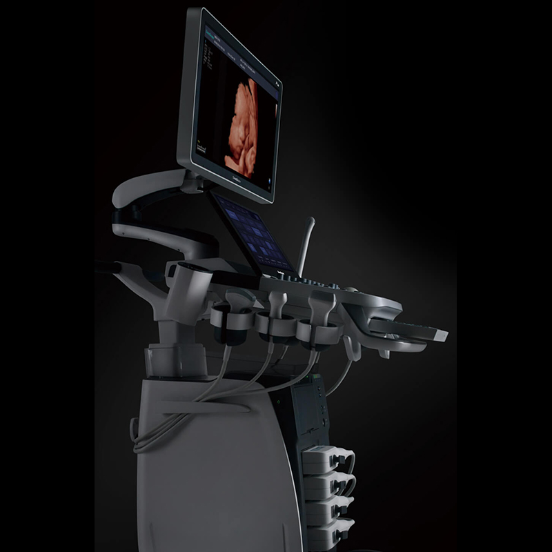
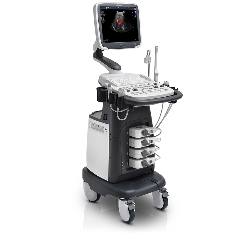
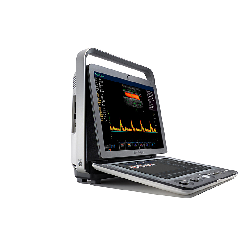
The P20 has many user-friendly designs, a simple control panel, an intuitive user interface, and a variety of automated auxiliary scanning functions, which can significantly improve daily work efficiency and help you quickly complete image acquisition and diagnosis. While taking into account the application of the whole body, P20 also has professional 4D function, making its application in the field of obstetrics and gynecology also handy.
Ultra-wideband system platform
The ultra-wide system platform inherited from Wi-Sono, combined with the advantages of ultra-wideband probe technology, can better obtain the best balance of high resolution and high penetration to ensure image quality.
μ-Scan micro imaging technology
Micron imaging technology adopts new algorithms to further enhance the processing ability of tissue structure information, and strive to provide more realistic diagnostic information for the clinic.
The elastic imaging function adopts updated software algorithms to easily obtain stable and reliable high-quality elastic images. At the same time, it is equipped with quantitative analysis software and unreliable area automatic elimination technology to provide you with a full range of diagnostic information.
Intelligent recognition of tissue information, automatic image splicing, can fully display the anatomical structure of larger lesions or tissues and organs, and cooperate with UHF linear array probe to provide professional solutions for musculoskeletal applications.
The position and angle of the sampling line can be adjusted arbitrarily for the anatomical M type, and more accurate heart function measurement data can be obtained by vertical measurement of the myocardial structure, which effectively evaluates myocardial movement and left ventricular function.
Tissue Doppler imaging can accurately obtain and visually display myocardial movement information, making it more objective and reliable to evaluate myocardial movement and function.
Auto EF Endocardial automatic tracing can automatically identify the systolic and end-diastolic endocardium, and automatically calculate the EF value.
Auto IMT can quickly obtain the inner media thickness of the anterior and posterior walls of the blood vessel, improving the repeatability of the measurement results and the inspection efficiency.
Auto Face fetal facial recognition can instantly remove fetal facial obstructions with one button.
The automatic volume measurement of AVC Follicle can identify and measure multiple follicles at the same time.
Auto NT automatically measures the transparent layer of the fetal neck can help doctors quickly and accurately obtain the NT value, reducing manual measurement errors.
AutoC intelligent blood flow tracking technology can automatically adjust the position and deflection angle of the ROI to help quickly obtain the best blood flow image.
Welcome to the website of Henan Anhel Medical Equipment Co., Ltd.!
- Molecular diagnosis
- Rapid Tests
- Drug of Abuse
-
Immunofluorescence
- Urinalysis Tests
-
POCT System
-
ELISA Test
-
DOT Test
-
Chemistry
- Ultrasonic
- Hemodialysis
- Ventilator
- ECG/EEG/EMG
-
Dermatology
- AN-specific electromagnetic wave therapy device
- AN-KL Carbon Dioxide Laser Treatment Machine (Scanning)
- AN high-energy narrow-band red and blue light therapy system
- AN Steam Therapy Fumigation Apparatus
- AN-308nm Excimer Light Skin Treatment System
- AN-5000 308 Home Excimer Phototherapy Apparatus
- AN-LED Home Spectral Therapy Apparatus
- AN 311 Ultraviolet Light Therapy Apparatus
- AN LED Spectrum Therapy Apparatus
- AN Medical Video Wood Lamp
- CT/MR
-
Ophthalmology Department
- AN-BLX5 Dental X-Ray
- AN-BLX10 Dental X-Ray
- AN-BLX8P High Frequency Dental X-Ray
- AN-BLX9 Dental X-Ray
- AN-BLX6 Dental X-Ray
- AN-BLX8 Dental X-Ray
- AN-digital oral X-ray imaging system HDR-500/600
- AN-Visual evoked potential meter M-800E
- AN-AOV-FB Excimer Laser Ophthalmology Treatment Machine
- AN-YZ3 Handheld Slit Lamp Microscope
- Dental
- Hematology Analyzer
- Chemistry Analyzer
-
POCT SYSTEM
- AN-AT1 Touch Screen HD Black and White Ultra
- AN-RS-N50 full digital notebook ultrasound diagnostic instru
- AN-RS-N50 (VET) all-digital notebook ultrasound diagnostic i
- AN-A6 All-digital veterinary B-ultrasound diagnostic instrum
- AN-M6 Veterinary B Ultrasound Diagnostic Instrument
- AN-A10 All-digital veterinary B-ultrasound diagnostic instru
- AN-M10 Veterinary B Ultrasound Diagnostic Instrument


