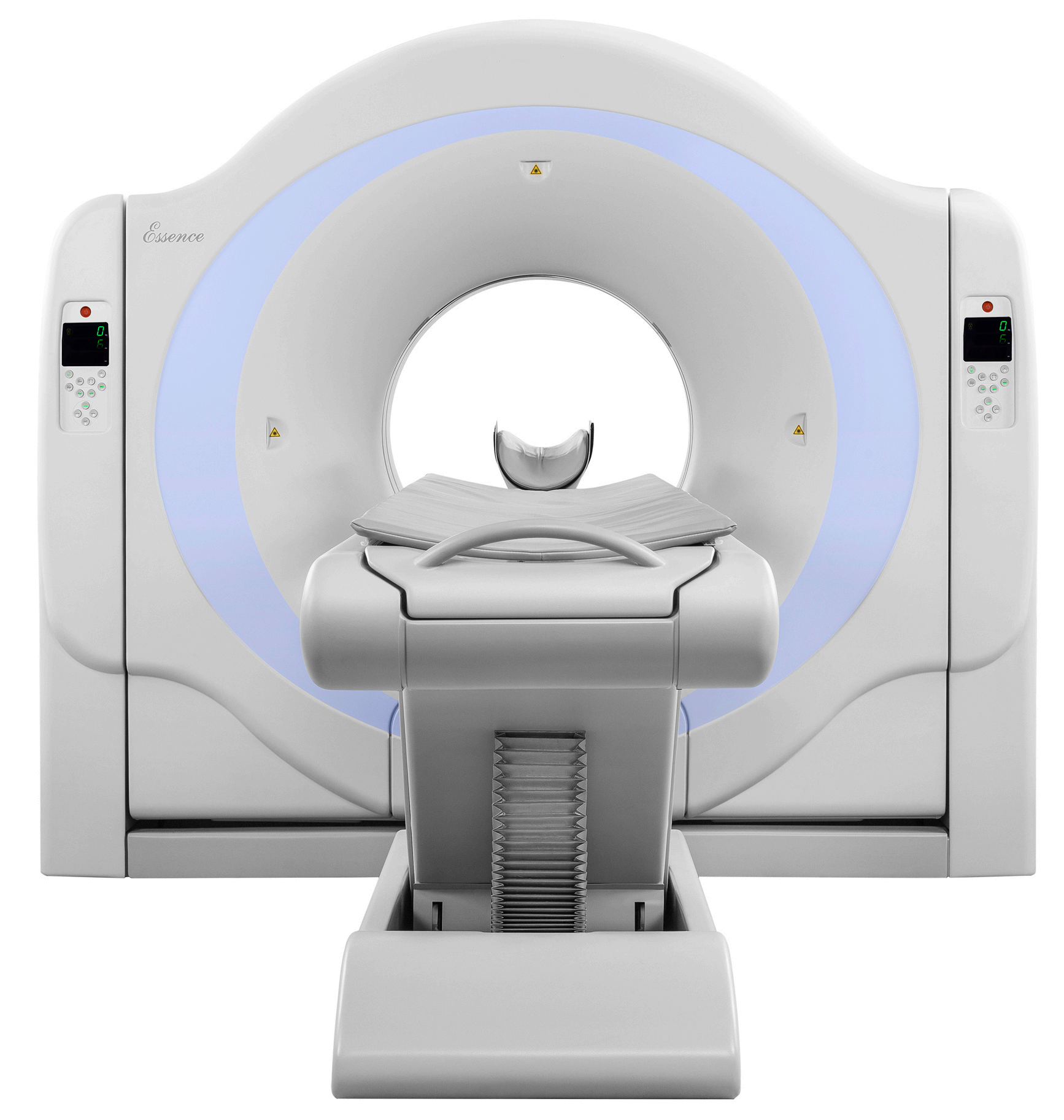
New Advanced Technology
Iterative algorithm, coronary artery analysis, fat
analysis and many more advanced applications, bring
patients more safety, all-sided clinical applications.
Hardware Configuration
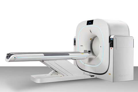
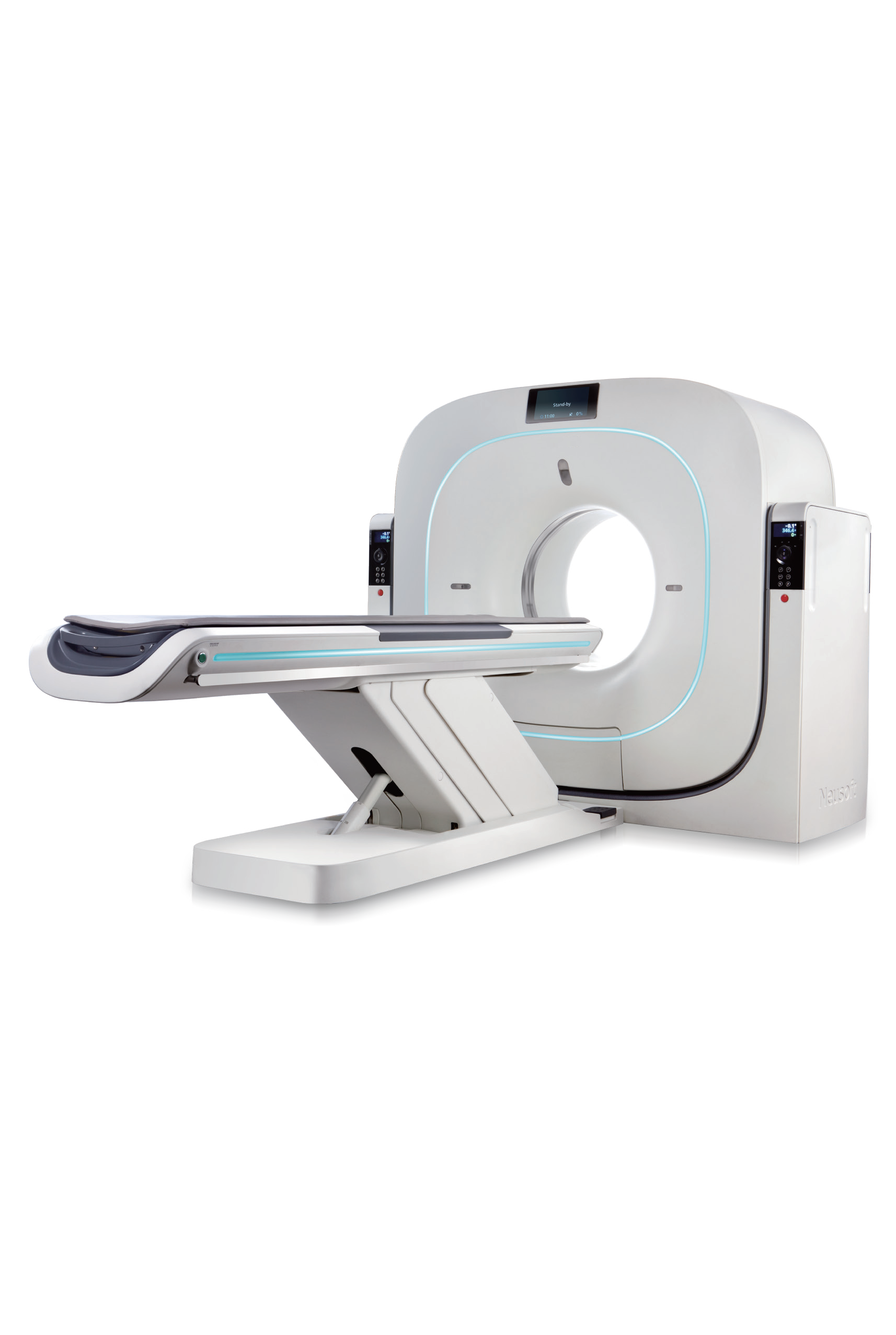
1.Gantry System
Aperture: 72 cm
Scan Field: 50 cm
Tilt: ± 30°
Rotation Time: 0.374s, 0.5s, 0.6s, 0.8s, 1.0s, 1.5s,
2.0s
Partial Scan Times (240°): 0.25s, 0.32s, 0.39s, 0.52s,
0.65s, 0.97s, 1.29s
Focus to Iso-center Distance: 570mm
Focus to detector Distance: 1040mm
Cooling Method: Air cooling
2.Data Acquisition System
Max. number of Slices: 128 Slices/Rotation
Number of Detector Rows: 64 rows
Number of Detector Elements:672X64
Total Channels per Slice: 1344
Min. Slice Thickness: 0.625mm
Detector Width: 40mm
Number of Projections: 4640
Detector Type: Solid State
Sequence Acquisition Modes: 128x0.625,
64x0.625, 32x0.625, 16x0.625, 8x0.625, 2x0.625
Spiral Acquisition Modes: 128x0.625, 64x0.625,
32x0.625, 16x0.625, 16x0.3125, 8x0.625 Detector:
Up to 50% SNR improvement compared to
conventional CT detectors;
Down to 1us-2us decay time for sub second scan
application;
Ultra low afterglow;
Special design to minimize electronic noise;
High geometric efficiency
3.X-ray Tube & Generator
Tube Current Range: 10mA~667mA
Tube Voltage: 80kV, 100kV, 120kV, 140kV
Tube Anode Heat Storage Capacity: 8.0MHU
Cooling Rate: 931kHU/min
High Tube Capacity high patient throughput
Focal Spot Size: 0.6×1.2mm(Small);
1.1×1.2mm(Large)
Max. Power: 80kW
Generator Type: High Frequency
4.Patient Table
Max. table Load: 205kg/452 lbs
Table Feed Speed: 1mm/s-160mm/s
Vertical Table/Travel Range: 430mm-970mm
Vertical Travel Speed: 9 mm/s-15mm/s
Movement Range: 1770mm
Table pad material: Carbon Fiber
5. Host Computer Systems
The host computer workplace provides an intelligent
and reliable workflow for data acquisition, image
reconstruction, and routine ppost processing at the CT
scanner.
Monitor: 24 inch, 1920 x 1200 resolution; Barco
Host: Dell Precision T5820
CPU: Intel Xeon W-2123 Processor (4 Core, 3.6GHz,
5.5MB Cache,8T)
RAM: 16GB (2 x 8GB, 2666MHz, DDR4, RDIMM)
GPU: NVIDIA Quadro P400, 2GB
System Disk: 1TB, 3.5 inch SATA3.0 Disk (7,200 rpm)
Data Disk: 1TB, 3.5 inch SATA3.0 Disk (7,200 rpm)
Recon: Dell T640
CPU: Intel Xeon Gold 6134 Processor (8 Core,
3.7GHz, 24.75MB Cache,16T)
RAM: 128GB (8 x 16GB, 2666MHz, DDR4, RDIMM)
GPU: 3 x LeadTek RTX 2060
System Disk: 1TB, 3.5 inch SATA3.0 Disk (7,200 rpm)
Data Disk: 4TB, 3.5 inch SATA3.0 Disk (7,200 rpm)
Images Additional Storage:
CD-R 700 MB 1,100 Images
DVD DICOM Drive 4.7 GB DVD Media 8,400 Images
Write-RW/+RW/-DL/Read
DICOM Viewer: Included on each CD;
Automatically started on the viewer's PC
6. AVW Workplace Systems
AVW workplace provides the unique advantage of an
efficient multi-modality diagnostic workflow at a single
workplace.It manages the clinical diagnostic workflow
anywhere within the clinical environment.
Monitor: 24 inch; 1920 x 1200 resolution; Beacon
Display HL2416SL
Computer Configuration: Dell Precision 3630
CPU: Intel Xeon E-2124G Processor (4 Core, 3.4GHz,
8MB Cache, 4T)
RAM: 16GB (2 x 8GB, 2666MHz, DDR4, UDIMM)
GPU: Leadtek GTX 1660Ti
System Disk: 256GB, 2.5 inch SATA3.0 Disk SSD
Data Disk: 1TB, 3.5 inch SATA3.0 Disk (7,200 rpm)
Additional Storage: CD-R 700 MB 1,100 Images DVD
DICOM Drive 4.7 GB DVD Media 8,400 Images Write�
RW/+RW/-DL/Read
DICOM Viewer:Included on each CD;
Automatically started on the viewer's PC
7. System Performance
Patient Registration: Direct input of patient information;
Acquisition Workplace immediately prior to scan;
Pre-registration of patients at any time prior to scan;
Special emergency patient registration (allows
examination without entering patient data before
scanning);
Transfer patient information from HIS/RIS via DICOM
Worklist;
Transfer examination information from scanner into
HIS/RIS via MPPS (Modality Performed Procedure
Step)
Up to 10,000 protocols can be edited, modified, and
stored, the doctors can modify and create the
protocols freely!
Surview
Length: 501700mm
Scan Times: 1.518 s
Views: A.P., Lateral, Dual
Real-Time Topogram: Yes
Sequence Acquisition
Reconstructed Slice Widths: 0.625, 1.25, 2.5, 5,
10mm Dynamic Multi-Scan: Multiple (continuous)
sequence scanning without table movement for fast
dynamic contrast studies with maximum slice
thickness of 40mm
Scan Length: Max. 1800mm
Multi-slice Spiral Acquisition
Reconstructed Slice Widths: 0.4, 0.625, 0.8, 1, 1.25,
1.5, 2, 2.5, 3, 4, 5, 6, 7, 8, 9,10mm
Slice Increment: 0.120 mm
Spiral Scan Time: Max. 100 s
Scan Length: Max. 1700mm
Pitch: 0.13-1.5
8.Image Reconstruction
Real-Time Display: Real-time image display during
spiral acquisition.
Scan Field: 50 cm
Recon Field: 550 cm
Recon Time: Up to 40 images/s with full cone beam
reconstruction
Recon Matrix: 512x512, 768x768,1024x1024
Display Matrix: 512x512, 768x768,1024x1024
HU Scale: 32,768 to +32,767
9.CINE Display
Display of Image Sequences
Automatic or Interactive with Mouse Control Max.
Image Rate: 30 frames/s
10.Filming
Connection via DICOM Basic Print Interactive Virtual
film Sheet
11.Image Transfer/Networking
Interface for transfer of medical images and
information using the DICOM 3.0 standard.
Facilitates communication with devices from different
manufactures.
DICOM Storage (Send/Receive)
DICOM Query/Retrieve
DICOM Basic print
DICOM Get Worklist (HIS/RIS)
DICOM MPPS
DICOM Storage Commitment
DICOM Viewer on CD
Raw Data Capacity: 2.4TB
12. Image Quality
Low-contrast Resolution
Object Size: 2mm@0.3%
Dose (CTDIw): 18mGy
High-contrast Resolution
Isotropic high-contrast resolution in all three planes
(x, y, and z).
X-Y-Plane
0%MTF 17lp/cm, 0.29mm
24lp/cm, 0.21mm, iHD
10%MTF 11lp/cm, 0.45mm
50%MTF 7.5lp/cm, 0.66mm
Z-Plane
0%MTF 15.0lp/cm, 0.33mm
10%MTF 10.0lp/cm, 0.5mm
50%MTF 6.0lp/cm, 0.83mm
Technique
245mA, 120kV, 1.0s, 0.625mm
Noise: <=0.35%
Artifact reduction
· Beam hardening compensation
· Metal artifact reduction
· Motion artifact reduction
· Volume artifact reduction
·
Adaptive streak Artifact reduction
· Lung intensification
· Advanced noise reduction


